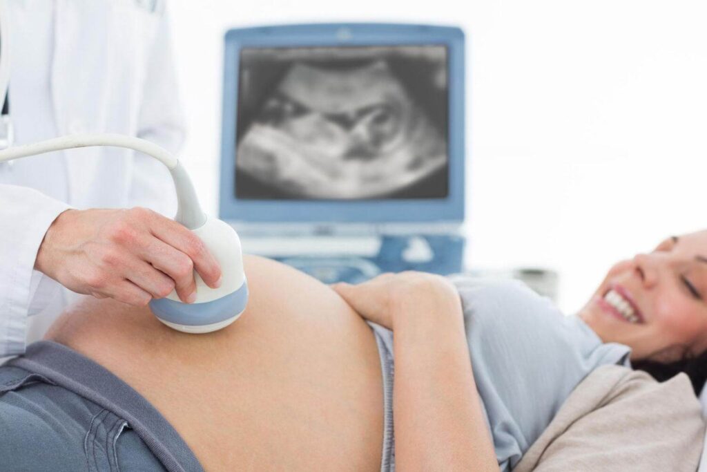
3D/4D Ultrasound Examination
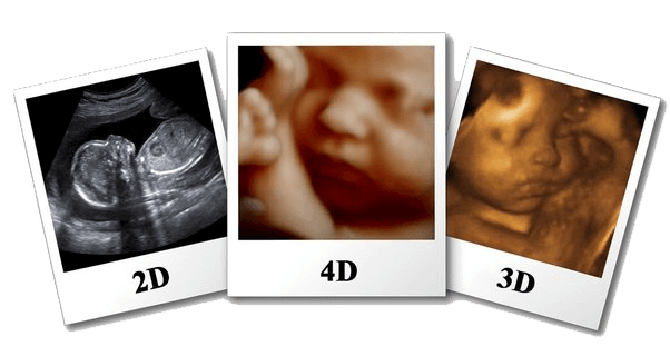
3D- is a static image of fetus in three dimensions (length, height and depth). 4D gives the possibility to receive third dimensional image of fetus in real time which means we are able to observe movement of fetal legs and hands, yawning, smile and so on in real time.
3D/4D examination is performed at any gestational age, but it should be taken into consideration that the smaller is gestational age, the better is fetal visualization and as the gestational age advances, the better images of face and its structures are obtained. Upon our recommendation conduction of 3D/4D examination is preferable at 26-32 weeks of pregnancy. The results of the examination are recorded on CD and DVD.
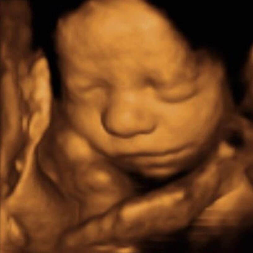
Three dimensional image is especially valuable to assess external abnormalities of fetus. With the help of 3D ultrasound physicians have an opportunity to make assessment of various fetal structures in thee projections that is of paramount importance to determine congenital anomalies. The information obtained via this examination is essential to define anomalies of extremities, face, vertebral column and other organs. With the help of 3D ultrasound examination determination of gender is more simplified.
The duration of this examination is 20-30 minutes. It is noteworthy that compared to an ordinary examination during 3D/4D ultrasound examination intensity and capacity value of ultrasound wave is not increased.
Positive aspects
3D/4D ultrasound examination has its positive aspects. The spacial images received during 3D/4D US examination give a chance to inspect fetal structures better compared to two dimensional images. Also, the image received is appreciated more easily by parents and other specialists.