


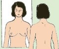
Self-examination is carried out in three positions:
 Self-examination is carried out in three positions:
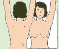
 While on your back, abduct an arm - "arms to the sides" position
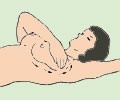
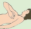 Being in dorsal position you should relax you hand along your body and examine breast by an opposite hand.
External half of mammary gland is examined by fingers of opposite hand. You start examination from nipple and move your fingers towards upper external quadrant. 

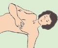
Then you examine all parts of internal side via the following order: you start examination from nipple and move your hand towards breast bone. You should look for lumps, what consistency they have, if they are painful.
Then you examine axillary and infraclavicular areas and nipple: apply pressure to nipple and areola gently by two fingers, watch if there is any discharge. 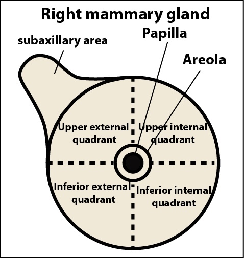
and nipples: gently hold the nipple and areola area with two fingers. Check for discharge.
Discharge may be:
• From one or both mammary glands;
• Spontaneous or released by pressure;
• Discharge maybe of different color: bloody, greenish, yellow or white.
It should be taken into account that in addition to cancer various reasons exist that can cause formation of mass in mammary gland and cause discharge from nipple.
Thus, if unordinary thing is noticed you have to visit a specialist (gynecologist, breast specialist) on time. Only he/she is able to make an accurate diagnosis based on special examinations.